
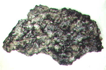
Terrestrial life (contaminate) on Martian meteorite Los Angeles
 |
 |
 |
|
 |
 |
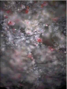
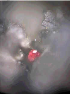
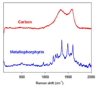
Red spots observed on the flat sawn surface of B side of the rock chip have a characteristic Raman spectrum of organic material (Fig. 3). The major peaks near 1361, 1490, 1556, and 1603 cm-1 match the characteristic I, II, III, and IV peaks of metalloporphyrin compounds [9]. The strong intensity of I, II, and IV peaks could arise from resonance Raman enhancement common in metalloporphyrins excited with a blue-green laser line. We also find a greater likelihood of detecting hematite in grains near red spots. | |
Reference: Wang A., Kuebler, K.E., Freeman J., Jolliff B. L., Preliminary Raman spectroscopic survey on a martian meteorite – Los Angeles, 32th LPSC, 2001.
![]()